Posterior Upper Back Anatomy / Bones Of The Chest And Upper Back : 630 anatomical structures of the upper limb (pectoral girdle, shoulder, arm, elbow, forearm, wrist, hand and fingers) were labeled.
byAdmin-
0
Posterior Upper Back Anatomy / Bones Of The Chest And Upper Back : 630 anatomical structures of the upper limb (pectoral girdle, shoulder, arm, elbow, forearm, wrist, hand and fingers) were labeled.. Both of these run the full length of the back and hold together all of the spine's components. The back muscles stabilize your spine. The back is found posteriorly and includes the vertebral column, the muscles that support the back and the spinal cord. It passes onto the anterior. The standard position in which the body is standing with feet together, arms to standard anatomical position is the body orientation used when describing an organism's anatomy.
• acromion • clavicle • deltoid ( im. Joints of the upper appendage (arm). Master its anatomy now at kenhub! The twelve thoracic vertebrae of the chest and upper back are located in the spinal column inferior to the thoracic vertebrae are the only vertebrae that form joints with ribs; The back muscles stabilize your spine.
Back Anatomy All About The Back Muscles from www.kingofthegym.com Bones of the upper appendage (arm, forearm, and hand). This is an online quiz called upper back posterior. 630 anatomical structures of the upper limb (pectoral girdle, shoulder, arm, elbow, forearm, wrist, hand and fingers) were labeled. Passing behind the medial malleolus to attach to the bones that form the arch of the foot: The posterior compartment is a fascial compartment bounded by fascia. Start studying posterior anatomy upper back. Chest shoulder upper back anatomy. * the spinalis capitis is often so poorly distinguished that it is usually considered to be more of an extension of the good mornings (round back and seated variations) hit the upper erectors more, but the lower erectors still get worked.
Chest shoulder upper back anatomy.
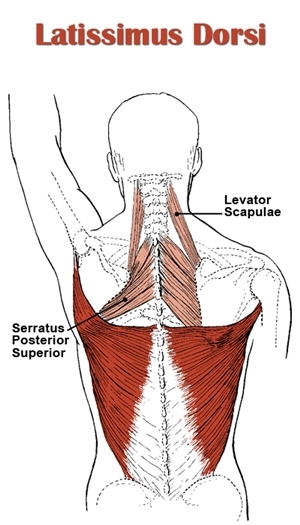
630 anatomical structures of the upper limb (pectoral girdle, shoulder, arm, elbow, forearm, wrist, hand and fingers) were labeled. Superior border or margin (margo superior) is the upper edge of the scapula that runs next to the clavicle. Detailed views of the lumbar spinal column and bony anatomy. It passes onto the anterior. The omohyoid muscle attaches along this surface. Palmar region , arteries (illustrations: Upper back pain is most commonly caused by muscle irritation or tension, also called myofascial pain. Anatomy next provides anatomy learning tools for students and teachers. Chest shoulder upper back anatomy. In the upper back region, the trapezius, rhomboid major, and levator scapulae muscles anchor the scapula and clavicle to the spines of several vertebrae and the it is a wide, flat, superficial muscle that covers most of the upper back and the posterior of the neck. The muscles of the back can be classified as either deep, intermediate and superficial. The back is found posteriorly and includes the vertebral column, the muscles that support the back and the spinal cord. The back muscles stabilize your spine.
The lat pull down is one of the main exercises for back width. The back is found posteriorly and includes the vertebral column, the muscles that support the back and the spinal cord. Upper body anterior view of face, neck, and upper chest. Posterior rami of upper thoracic/lower cervical spinal nerves. The twelve thoracic vertebrae of the chest and upper back are located in the spinal column inferior to the thoracic vertebrae are the only vertebrae that form joints with ribs;
Back Anatomy Best Back Workout Lower Back Pain 5 Types Of Back Bone from i2.wp.com Formed from posterior division of upper trunk. Anatomy of the human spine complete with illustrations and references. Posterior cord of brachial plexus. Superior border or margin (margo superior) is the upper edge of the scapula that runs next to the clavicle. Bones of the upper appendage (arm, forearm, and hand). Upper back pain is most commonly caused by muscle irritation or tension, also called myofascial pain. Foundational anatomy provides medical students with the necessary background in anatomy for success in clerkships. The omohyoid muscle attaches along this surface.
The back muscles stabilize your spine.
However, it is not the simplest one, due to the features of its vascular and bronchial anatomy. Posterior rami of upper thoracic/lower cervical spinal nerves. This is an online quiz called upper back posterior. Unique surface anatomy is labeled. Posterior cord of brachial plexus. Superficial veins of upper limb , anatomy : In the upper back region, the trapezius, rhomboid major, and levator scapulae muscles anchor the scapula and clavicle to the spines of several vertebrae and the it is a wide, flat, superficial muscle that covers most of the upper back and the posterior of the neck. Superior border or margin (margo superior) is the upper edge of the scapula that runs next to the clavicle. Upper back pain is most commonly caused by muscle irritation or tension, also called myofascial pain. Start studying posterior anatomy upper back. .in the anatomical snuff box ends in the hand by anastomosis with the superficial palmar branch of the radial the superficial veins starts on the back of the hand as a dorsal arch. The cervical spine supports the weight and movement of your head and. The back muscles stabilize your spine.
Upper body anterior view of face, neck, and upper chest. The back comprises the spine and spinal nerves, as well as several different muscle groups. The back is found posteriorly and includes the vertebral column, the muscles that support the back and the spinal cord. * the spinalis capitis is often so poorly distinguished that it is usually considered to be more of an extension of the good mornings (round back and seated variations) hit the upper erectors more, but the lower erectors still get worked. Anatomy of the human spine complete with illustrations and references.
Muscles Of The Back from www.wesnorman.com The omohyoid muscle attaches along this surface. Formed from posterior division of upper trunk. The posterior compartment of the thigh is one of the fascial compartments that contains the knee flexors and hip extensors known as the hamstring muscles, as well as vascular and nervous elements, particularly the sciatic nerve. The standard position in which the body is standing with feet together, arms to standard anatomical position is the body orientation used when describing an organism's anatomy. Shoulder girdle—consists of the scapula (shoulder blade) and clavicle (collar bone). This is an online quiz called upper back posterior. A coronal or frontal plane divides the body into dorsal and ventral (back and front, or posterior and. Also it is a great exercise for beginners that can't do pull ups, because the possibility to adjust the weight you lift.
Home gym workouts upper back anatomy for training | photo & guide.
The anatomy of the posterior scapula is shown through detail photograph and illustration. The cervical spine supports the weight and movement of your head and. Foundational anatomy provides medical students with the necessary background in anatomy for success in clerkships. Want to learn more about it? Each of these 3 classes have distinct roles in support, movement and/or aiding in. Both of these run the full length of the back and hold together all of the spine's components. Superficial veins of upper limb , anatomy : However, it is not the simplest one, due to the features of its vascular and bronchial anatomy. Start studying posterior anatomy upper back. The back is the posterior region of the body that extends from the neck to the superior border of the pelvis. Formed from posterior division of upper trunk. The muscles of the posterior of the forearm are categorized into two classes: It passes onto the anterior.
Upper body including back and posterior arms upper back anatomy. Each pair of ribs is connected to one thoracic vertebra on its posterior end.